
How We See What You Can't: Eye Technology Explained
OPTOMAP
Medical Perspective: Ultra-widefield retinal imaging (Optomap) captures up to 200° of the retina in a single image without dilation, allowing screening for peripheral pathology including retinal tears, holes, lattice degeneration, and early melanomas that may be asymptomatic but vision-threatening if undetected.
Everyday Perspective: Think of an Optomap like your car's state inspection - a comprehensive "once-over" to make sure nothing critical is being overlooked. We can see about 80% of your retina in seconds, catching problems in the far edges of your eye that you'd never notice until they threaten your central vision. It's preventive screening that finds issues before they become emergencies.
OPTIC NERVE
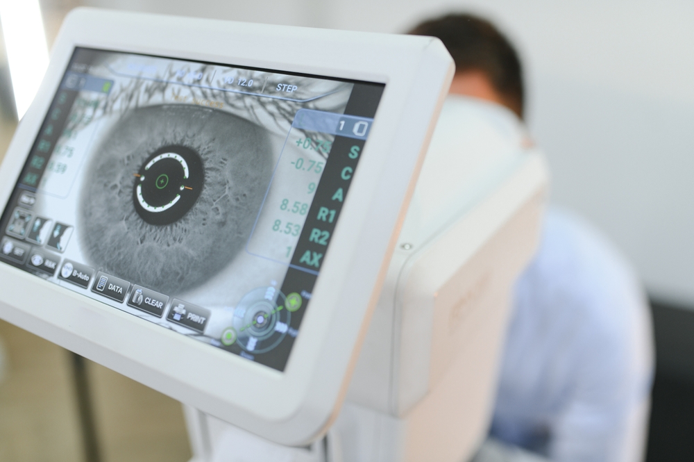
Medical Perspective: The optic nerve contains approximately 1.2 million retinal ganglion cell axons that transmit visual information from photoreceptors to the lateral geniculate nucleus and visual cortex. Any disruption along this pathway - whether at the nerve head, orbital portion, or chiasm - results in corresponding visual field defects.
Everyday Perspective: Your eyeball connects to your brain through the optic nerve - think of it like an HDMI cable connecting your computer to a monitor. The eye captures the image, but the nerve transmits that signal to your brain where vision actually happens. Damage anywhere along that cable causes picture problems, even if the camera (eye) and monitor (brain) are working fine.
VISUAL FIELDS
Medical Perspective: Visual field testing maps functional deficits along the entire visual pathway from retina through optic nerve, chiasm, optic tracts, and visual cortex. While fundoscopy reveals structural optic nerve damage, perimetry detects functional losses that localize lesions anywhere along this pathway - including retrobulbar, chiasmal, or post-chiasmal pathology invisible on eye examination.
Everyday Perspective: During an eye exam, I can only see where the optic nerve enters your eye - like seeing where electrical wiring enters the wall. Visual field testing lets me see how that "wiring" is functioning all the way through the wall to the brain. If electricity isn't getting where it should be, this test shows me whether the problem is at the outlet (eye), in the wall (nerve pathway), or at the breaker box (brain).

MAP
Contact Info
Hours of Operation
- Monday 10:00am - 7:00pm
- Tuesday 8:00am - 4:30pm
- Wednesday 8:00am - 4:30pm
- Thursday 8:00am - 4:30pm
- Friday 7:30am - 1:00pm
- Saturday Closed
- Sunday Closed
© 2026 Everett Eye Care Center & Med Spa. All rights Reserved. Accessibility Statement - Privacy Policy - Sitemap
Powered by: